
Sunday, September 11, 2011
Thursday, August 4, 2011
MOLECULAR GENETICS OF DEVELOPMENT AND CELL DIFFERENTIATION IN MOUSE AND MAN

The development of complex organs and tissues, such as brain and the hematopoietic system, requires the ordered expression of key transcription factors controlling cell type- and tissue-specific gene expression. Stem cells represent the self renewing compartment of rapidly replicating cell types, as in the hematopoietic system, but are present, in small numbers, also in adult brain, heart and other organs which do not show active cell replication in adults.
The group uses a common set of approaches (conditional and standard targeted mutagenesis in mouse, cell culture and gene transduction, chromatin studies, etc.) to investigate the role of key transcription factors in the development, maintenance and differentiation of a variety of stem cells.
Molecular Computing

Molecular computation is an emerging area of study as a borderline between computational science, chemistry and biology. The objective of this study is to create an information processing mechanism using chemical reactions of biomolecules such as DNA. Other objectives include designing biomolecules and controlling their chemical reactions using information technology. Application of computational science to chemistry and biology will be discussed in this course through explanation of molecular computation and its related fields.
Privacy Policy
Privacy Policy for http://molecular-and-cell.blogspot.com/
If you require any more information or have any questions about our privacy policy, please feel free to contact us by email at joko.kentir22@gmail.com.
At http://molecular-and-cell.blogspot.com/, the privacy of our visitors is of extreme importance to us. This privacy policy document outlines the types of personal information is received and collected by http://molecular-and-cell.blogspot.com/ and how it is used.
Log Files
Like many other Web sites, http://molecular-and-cell.blogspot.com/ makes use of log files. The information inside the log files includes internet protocol ( IP ) addresses, type of browser, Internet Service Provider ( ISP ), date/time stamp, referring/exit pages, and number of clicks to analyze trends, administer the site, track user’s movement around the site, and gather demographic information. IP addresses, and other such information are not linked to any information that is personally identifiable.
Cookies and Web Beacons
http://molecular-and-cell.blogspot.com/ does use cookies to store information about visitors preferences, record user-specific information on which pages the user access or visit, customize Web page content based on visitors browser type or other information that the visitor sends via their browser.
DoubleClick DART Cookie
.:: Google, as a third party vendor, uses cookies to serve ads on http://molecular-and-cell.blogspot.com/.
.:: Google's use of the DART cookie enables it to serve ads to users based on their visit to http://molecular-and-cell.blogspot.com/ and other sites on the Internet.
.:: Users may opt out of the use of the DART cookie by visiting the Google ad and content network privacy policy at the following URL - http://www.google.com/privacy_ads.html
Some of our advertising partners may use cookies and web beacons on our site. Our advertising partners include ....
Google Adsense
These third-party ad servers or ad networks use technology to the advertisements and links that appear on http://molecular-and-cell.blogspot.com/ send directly to your browsers. They automatically receive your IP address when this occurs. Other technologies ( such as cookies, JavaScript, or Web Beacons ) may also be used by the third-party ad networks to measure the effectiveness of their advertisements and / or to personalize the advertising content that you see.
http://molecular-and-cell.blogspot.com/ has no access to or control over these cookies that are used by third-party advertisers.
You should consult the respective privacy policies of these third-party ad servers for more detailed information on their practices as well as for instructions about how to opt-out of certain practices. http://molecular-and-cell.blogspot.com/'s privacy policy does not apply to, and we cannot control the activities of, such other advertisers or web sites.
If you wish to disable cookies, you may do so through your individual browser options. More detailed information about cookie management with specific web browsers can be found at the browsers' respective websites.
Saturday, March 5, 2011
Basic extraordinary cell biology
During a recent visit with my 12-year old daughter’s science teacher, I mentioned that I had read a few books on cell biology over the past couple of years and that I was interested in sitting in on one of the upcoming sixth grade science classes–my daughter had mentioned that they were beginning to study cell biology. I mentioned a few of the things that I had found interesting about cells to the science teacher. After noticing my enthusiasm, she retracted her invitation to watch the class and, instead, invited me to teach part of the class. A few days later I made my science teaching debut.
I advised the sixth-graders that although I work as a lawyer during the day, I often read science books, and I often write about science on my website. I told them that I had no serious science education at the Catholic grade school I attended. I didn’t have any biology class at all until I was a sophomore in high school. That was mostly a nuts and bolts class taught by a Catholic nun who failed show the excitement the subject deserved. She also forgot to teach by Theodosius Dobzhansky’s maxim that “nothing in biology makes sense except in the light of evolution.”
I told “my” class that anyone who studies cells with any care will be greatly rewarded. Studying cells is actually autobiographical because “you are made of 60 trillion of cells.” These cells are so small that people cannot even see them.
One of the students then confused trillions for millions. “Keep in mind,” I cautioned, “that a trillion is a million million.” With regard to their size, there is only one human cell–the human ovum–that you can see with the naked eye—it is much bigger than the other cells in your body. Despite its tiny size, the human ovum is so incredibly small that it’s smaller than the period at the end of this sentence. See this wonderful illustration of the size of human cells, and many other small objects.
The volume of a eukaryotic cell is typically 1000 times larger than that of a prokaryotic one.
Page 28
Page 28
I told the students that the study of cells is autobiographical “because each of you is a community of cells. You are a self-organized community.” Even the brain is made of cells. It thinks, even though individual cells don’t think. Individual cells can’t think, but you can think. “How is that for amazing?” One girl raised her hand.
“I don’t understand how this can be. I don’t understand how the body can be made of trillions of cells. How can it possibly work? I have a lot of questions.”
I told her that her questions prove that she “gets it.” Truly, how can something as complex as a human body, or even as complex as a single cell possibly work? It’s amazing that these things work, yet most people more often focus on the times that they break down through disease or aging.
A bacterial cell consists of more than 300 million molecules (not counting water), several thousand different kinds of molecules, and requires some 2000 genes for specification. There is nothing random about this assemblage, which reproduces itself with constant composition and form generation after generation.
Page 10
Page 10
I didn’t claim to have many answers, but I told the students that I was there to share information I learned from my readings. I assured them that studying cells, including human cells, is more amazing than any fictitious story that they had ever read. Part of the reason the study of cells is so amazing is due to the complex anatomy of cells, especially eukaryotic cells. Appreciating much of the magic requires statistics. Some of it comes from the exquisite complexity of individual cells, however, and much of the magic derives from the appreciation that the scientific facts relating to cell biology are somehow true.

I then noticed a few of the students were looking puzzled. I reminded them that the scientific study of cells is not about trust. I was not asking them to trust me or their teacher. In upcoming classes, they will be invited to look into microscopes and see cells, including their own cheek cells or skin cells. With powerful microscopes we can even see chromosomes. I urged them to investigate more about cells on their own, because there is a wealth of information on the Internet. Go out there and check the evidence; investigate as skeptics. Believe only what you see. That’s what I did, and that’s why I’m excited to learn about cells. And remember that only 400 years ago, no one had any idea that humans were communities of cells. They are privileged to be living in an age where we have such detailed knowledge available to us.
I told the students that the information I would tell them came from a variety of sources, including a book called The Way of the Cell: Molecules, Organisms and the Order of Life, by Franklin M Harold (2001). I’ve inserted several passages from Franklin’s excellent book within this post. In case it isn’t apparent, this post is a summary of the sorts of things I taught my students. I found myself bouncing around the classroom fielding comments and questions and having a great time. My hope was that a few of the kids might see the subject of cell biology in a more compelling way after seeing me so revved about it. That was my main aim, to share my excitement.
Algorithms in Structural Molecular Biology and Proteomics
Some of the most challenging and influential opportunities for Physical Geometric Algorithms (PGA) arise in developing and applying information technology to understand the molecular machinery of the cell. Our recent work (e.g., [1-20]) shows that many PGA techniques may be fruitfully applied to the challenges of computational molecular biology. PGA research may lead to computer systems and algorithms that are useful in structural molecular biology, proteomics, and rational drug design.

Concomitantly, a wealth of interesting computational problems arise in proposed methods for discovering new pharmaceuticals. I'll briefly discuss some recent results from my lab, including new algorithms for interpreting X-ray crystallography [14, 17, 16] and NMR (nuclear magnetic resonance) data [3,9,6,19,10,5,7,18,4], disease classification using mass spectrometry of human serum [12], and protein redesign [13]. Our algorithms have recently been used, respectively, to reveal the enzymatic architecture of organisms high on the CDC bioterrorism watch-list [17,16], for probabilistic cancer classification from human peripheral blood [12], and to redesign an antibiotic-producing enzyme to adenylate a novel substrate [13]. I'll overview these projects, and highlight some of the algorithmic and computational challenges.
Toxicogenomics and Molecular Biology

Toxic tort claims, health risk assessments, biomonitoring, crime-scene investigations, and intellectual property cases are increasingly relying on techniques that fall under the realm of molecular biology-based science or "Toxicogenomics." Staying abreast of the rapidly evolving, cutting-edge science in this field is essential to understanding and interpreting health claims and in assessing health risks when molecular biology techniques are being used. Exponent scientists have experience in evaluating and conducting studies in the field of molecular biology, including genomics approaches such as DNA array (gene chip), real-time Q-PCR, cloning, transgenics, stem-cell therapeutics, RNA anti-sense, knock-outs, and knock-downs; and proteomics approaches such as, recombinant protein expression, cytokine assays, ELISA, radioisotope and immuno-labeling, and protein purification and identification through 1 and 2-D electrophoresis, and column chromatography.
Increasingly, "Gene Array" or DNA profiling and Cytokine arrays are being applied in an effort to understand disease mechanisms at the molecular level. In toxic tort claims, this technology is also being exploited in an to attempt to assign genetic profiles, "footprints," or "molecular signatures” to various chemical exposure scenarios, such as benzene and asbestos in order to validate an injury claim. In many cases there are considerable limitations to the design and interpretation of the scientific studies being used to support these claims. Our strong understanding of biochemistry, molecular interactions, and fate of chemicals in the body ensures a thorough scientific evaluation of molecular biology based approaches used to identify and quantify exposure and potential health effects. As part of an overall review of the science supporting a claim, Exponent scientists can critically evaluate the relevance of molecular mechanisms and techniques that are being invoked in a selected scenario.
As understanding of the molecular mechanisms behind toxicity evolves, government agencies are re-evaluating previously derived regulatory limits, and considering molecular biology-based data in deriving regulatory limits for new chemical compounds. Understanding the new molecular biology–based technology used to evaluate and assess risk of exposure to these chemicals is essential for conducting a weight-of-evidence review to determine new regulatory standards. As biotechnology patent applications increase, the need for third-party expert review is important for maintaining protection of intellectual property and "Freedom to Operate" in commercial research and development programs. Exponent scientists with experience both in the new molecular biology techniques and in regulatory toxicology can evaluate the science that will be used by regulatory agencies to derive these new limits and standards for patent protection.
As understanding of the molecular mechanisms behind toxicity evolves, government agencies are re-evaluating previously derived regulatory limits, and considering molecular biology-based data in deriving regulatory limits for new chemical compounds. Understanding the new molecular biology–based technology used to evaluate and assess risk of exposure to these chemicals is essential for conducting a weight-of-evidence review to determine new regulatory standards. As biotechnology patent applications increase, the need for third-party expert review is important for maintaining protection of intellectual property and "Freedom to Operate" in commercial research and development programs. Exponent scientists with experience both in the new molecular biology techniques and in regulatory toxicology can evaluate the science that will be used by regulatory agencies to derive these new limits and standards for patent protection.
Regulatory agencies and expert panels of scientists are recommending research and development of tools in the emerging field of "Toxicogenomics" for use in exposure assessment. A National Research Council committee on biomonitoring for environmental chemicals recently recommended the use of molecular biology-based biomonitoring approaches, in order "… to move beyond the traditional approaches of exposure assessment-based on one exposure to one chemical in one environmental medium" to assess multiple exposures and multiple biologic-response pathways using genetic markers of exposure and response.
Exponent has Ph.D.-level scientists who are widely published in the fields of genomics, proteomics, molecular biology, and biochemistry, with experience in designing, executing, and evaluating molecular biology-based studies. Let our scientists assist you in this rapidly evolving and complex field.
Cell Division and DNA Replication
In the first lecture, we covered the way science works and especially how the scientific method applies to biology. Then, we looked at the structure of the cell, building a map of the cell - knowing what processes happen where in the cell, e.g., the production of energy-rich ATP molecules in the mitochondria.
In the third part of the lecture, we took a closer look at the way DNA code gets transcribed into RNA in the nucleus, and the RNA code translated into protein structure in the rough endoplasmatic reticulum. Finally, we looked at several different ways that cells communicate with each other and with the environment, thus modifying cell function.
All of that information will be important in this lecture, as we cover the ways cells divide, how cell-division, starting with a fertilized cell, builds an embryo, how genetic code (genotype) influences the observable and measurable traits (phenotype) and, finally, how do these processes affect the genetic composition of the populations of organisms of the same species - the process of evolution.
Mitosis
The only way to build a cell is by dividing an existing cell into two. As the genome (the complete sequence of the DNA) is an essential part of a cell, it is neccessary for the DNA to be duplicated prior to cell division.
In Eukaryotic cells, chromosomes are structures composed mostly of DNA and protein. DNA is a long double-stranded chain-like molecule. Some portions of the DNA are permanently coiled and covered with protective proteins to prevent DNA expression (transcription). Other parts can be unraveled so transcription can occur.
The number of chromosomes is different in different species. Human cells possess 23 pairs of chromosomes. Prior to cell division each chromosome replicates producing two identical sister chromosomes - each eventually landing in one of the daughter cells.
The process of DNA replication - the way all of the DNA code of the mother cell duplicates and one copy goes into each daughter cell - is the most important aspect of cell division. It is wonderfully described in your handout and depicted in the animation. Other cell organelles also divide and split into two daughter cells. Once the process of DNA replication is over, the new portion of the cell membrane gets built transecting the cell and dividing all the genetic material into two cellular compartments, leading the cell to split into two cells.

Meiosis
Meiosis is a special case of cell division. While mitosis results in division of all types of cells in the body, meiosis results in the formation of sex cells - the gametes: eggs and sperm. Mitosis is a one-step process: one cell divides into two. Meiosis is a two-step process: one cell divides into two, then each daughter immediately divides again into two, resulting in four grand-daughter cells.
Each cell in the body has two copies of the entire DNA - one copy received from the mother, the other from the father. Fertilization (fusion of an egg and a sperm) would double the chromosome number in each generation if the egg and sperm cells had the duplicate copy. Meiosis ensures that gametes have only one copy of the genome - a mix of maternal and paternal sequences. Such a cell is called a haploid cell.
Once the egg and a sperm fuse, the resulting zygote (fertilized egg) again contains double dose of the DNA and is called a diploid cell. Thus the resultant zygote inherits genetic material from both its father and its mother. All the cells in the body except for the gametes are diploid. Sexual reproduction produces offspring that are genetically different from either parent.
DNA Replication
DNA replication is a complex process of duplication of the DNA involving many enzymes. It is the first and the most important process in cell division. Please read the handout (BREAKFAST OF CHAMPIONS DOES REPLICATION by David Ng) to appreciate the complexity of the process, but you do not need to memorize any of the enzymes for the exams. Also, it will help your understanding of the process if you watch this animation.
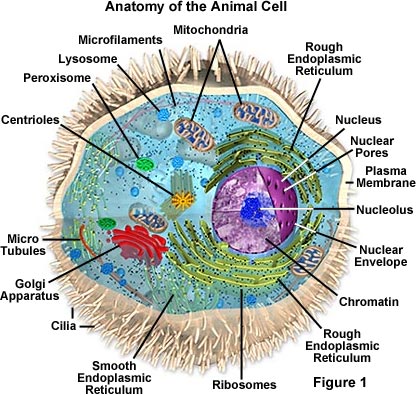
Animal Cell Structure
Animal cells are typical of the eukaryotic cell, enclosed by a plasma membrane and containing a membrane-bound nucleus and organelles. Unlike the eukaryotic cells of plants and fungi, animal cells do not have a cell wall. This feature was lost in the distant past by the single-celled organisms that gave rise to the kingdom Animalia. Most cells, both animal and plant, range in size between 1 and 100 micrometers and are thus visible only with the aid of a microscope.
The lack of a rigid cell wall allowed animals to develop a greater diversity of cell types, tissues, and organs. Specialized cells that formed nerves and muscles—tissues impossible for plants to evolve—gave these organisms mobility. The ability to move about by the use of specialized muscle tissues is a hallmark of the animal world, though a few animals, primarily sponges, do not possess differentiated tissues. Notably, protozoans locomote, but it is only via nonmuscular means, in effect, using cilia, flagella, and pseudopodia.
The animal kingdom is unique among eukaryotic organisms because most animal tissues are bound together in an extracellular matrix by a triple helix of protein known as collagen. Plant and fungal cells are bound together in tissues or aggregations by other molecules, such as pectin. The fact that no other organisms utilize collagen in this manner is one of the indications that all animals arose from a common unicellular ancestor. Bones, shells, spicules, and other hardened structures are formed when the collagen-containing extracellular matrix between animal cells becomes calcified.
Animals are a large and incredibly diverse group of organisms. Making up about three-quarters of the species on Earth, they run the gamut from corals and jellyfish to ants, whales, elephants, and, of course, humans. Being mobile has given animals, which are capable of sensing and responding to their environment, the flexibility to adopt many different modes of feeding, defense, and reproduction. Unlike plants, however, animals are unable to manufacture their own food, and therefore, are always directly or indirectly dependent on plant life.
Most animal cells are diploid, meaning that their chromosomes exist in homologous pairs. Different chromosomal ploidies are also, however, known to occasionally occur. The proliferation of animal cells occurs in a variety of ways. In instances of sexual reproduction, the cellular process of meiosis is first necessary so that haploid daughter cells, or gametes, can be produced. Two haploid cells then fuse to form a diploid zygote, which develops into a new organism as its cells divide and multiply.
The earliest fossil evidence of animals dates from the Vendian Period (650 to 544 million years ago), with coelenterate-type creatures that left traces of their soft bodies in shallow-water sediments. The first mass extinction ended that period, but during the Cambrian Period which followed, an explosion of new forms began the evolutionary radiation that produced most of the major groups, or phyla, known today. Vertebrates (animals with backbones) are not known to have occurred until the early Ordovician Period (505 to 438 million years ago).
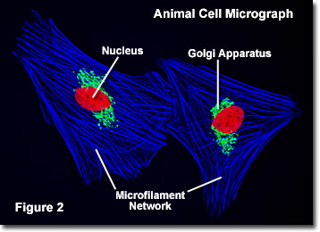
Cells were discovered in 1665 by British scientist Robert Hooke who first observed them in his crude (by today's standards) seventeenth century optical microscope. In fact, Hooke coined the term "cell", in a biological context, when he described the microscopic structure of cork like a tiny, bare room or monk's cell. Illustrated in Figure 2 are a pair of fibroblast deer skin cells that have been labeled with fluorescent probes and photographed in the microscope to reveal their internal structure. The nuclei are stained with a red probe, while the Golgi apparatus and microfilament actin network are stained green and blue, respectively. The microscope has been a fundamental tool in the field of cell biology and is often used to observe living cells in culture. Use the links below to obtain more detailed information about the various components that are found in animal cells.
- Centrioles - Centrioles are self-replicating organelles made up of nine bundles of microtubules and are found only in animal cells. They appear to help in organizing cell division, but aren't essential to the process.
- Cilia and Flagella - For single-celled eukaryotes, cilia and flagella are essential for the locomotion of individual organisms. In multicellular organisms, cilia function to move fluid or materials past an immobile cell as well as moving a cell or group of cells.
- Endoplasmic Reticulum - The endoplasmic reticulum is a network of sacs that manufactures, processes, and transports chemical compounds for use inside and outside of the cell. It is connected to the double-layered nuclear envelope, providing a pipeline between the nucleus and the cytoplasm.
- Endosomes and Endocytosis - Endosomes are membrane-bound vesicles, formed via a complex family of processes collectively known as endocytosis, and found in the cytoplasm of virtually every animal cell. The basic mechanism of endocytosis is the reverse of what occurs during exocytosis or cellular secretion. It involves the invagination (folding inward) of a cell's plasma membrane to surround macromolecules or other matter diffusing through the extracellular fluid.
- Golgi Apparatus - The Golgi apparatus is the distribution and shipping department for the cell's chemical products. It modifies proteins and fats built in the endoplasmic reticulum and prepares them for export to the outside of the cell.
- Intermediate Filaments - Intermediate filaments are a very broad class of fibrous proteins that play an important role as both structural and functional elements of the cytoskeleton. Ranging in size from 8 to 12 nanometers, intermediate filaments function as tension-bearing elements to help maintain cell shape and rigidity.
- Lysosomes - The main function of these microbodies is digestion. Lysosomes break down cellular waste products and debris from outside the cell into simple compounds, which are transferred to the cytoplasm as new cell-building materials.
- Microfilaments - Microfilaments are solid rods made of globular proteins called actin. These filaments are primarily structural in function and are an important component of the cytoskeleton.
- Microtubules - These straight, hollow cylinders are found throughout the cytoplasm of all eukaryotic cells (prokaryotes don't have them) and carry out a variety of functions, ranging from transport to structural support.
- Mitochondria - Mitochondria are oblong shaped organelles that are found in the cytoplasm of every eukaryotic cell. In the animal cell, they are the main power generators, converting oxygen and nutrients into energy.
- Nucleus - The nucleus is a highly specialized organelle that serves as the information processing and administrative center of the cell. This organelle has two major functions: it stores the cell's hereditary material, or DNA, and it coordinates the cell's activities, which include growth, intermediary metabolism, protein synthesis, and reproduction (cell division).
- Peroxisomes - Microbodies are a diverse group of organelles that are found in the cytoplasm, roughly spherical and bound by a single membrane. There are several types of microbodies but peroxisomes are the most common.
- Plasma Membrane - All living cells have a plasma membrane that encloses their contents. In prokaryotes, the membrane is the inner layer of protection surrounded by a rigid cell wall. Eukaryotic animal cells have only the membrane to contain and protect their contents. These membranes also regulate the passage of molecules in and out of the cells.
- Ribosomes - All living cells contain ribosomes, tiny organelles composed of approximately 60 percent RNA and 40 percent protein. In eukaryotes, ribosomes are made of four strands of RNA. In prokaryotes, they consist of three strands of RNA.
In addition the optical and electron microscope, scientists are able to use a number of other techniques to probe the mysteries of the animal cell. Cells can be disassembled by chemical methods and their individual organelles and macromolecules isolated for study. The process of cell fractionation enables the scientist to prepare specific components, the mitochondria for example, in large quantities for investigations of their composition and functions. Using this approach, cell biologists have been able to assign various functions to specific locations within the cell. However, the era of fluorescent proteins has brought microscopy to the forefront of biology by enabling scientists to target living cells with highly localized probes for studies that don't interfere with the delicate balance of life processes.
The Cell-Cell Structure

Life is both wonderful and majestic. Yet for all of its majesty, all organisms are composed of the fundamental unit of life, the cell. The cell is the simplest unit of matter that is alive. From the unicellular bacteria to multicellular animals, the cell is one of the basic organizational principles of biology. Let's look at some of the components of this basic organizer of living organisms.
Eukaryotic Cells and Prokaryotic Cells
There are two primary types of cells: eukaryotic cells and prokaryotic cells. Eukaryotic cells are called so because they have a true nucleus. The nucleus, which houses DNA, is contained within a membrane and separated from other cellular structures. Prokaryotic cells however have no true nucleus. DNA in a prokaryotic cell is not separated from the rest of the cell but coiled up in a region called the nucleoid.
As organized in the Three Domain System, prokaryotes include archaeans and bacteria. Eukaryotes include animals, plants, fungi and protists. Typically, eukaryoitc cells are more complex and much larger than prokaryotic cells. On average, prokaryotic cells are about 10 times smaller in diameter than eukaryotic cells.
As organized in the Three Domain System, prokaryotes include archaeans and bacteria. Eukaryotes include animals, plants, fungi and protists. Typically, eukaryoitc cells are more complex and much larger than prokaryotic cells. On average, prokaryotic cells are about 10 times smaller in diameter than eukaryotic cells.
Eukaryotes grow and reproduce through a process called mitosis. In organisms that also reproduce sexually, the reproductive cells are produced by a type of cell division called meiosis. Most prokaryotes reproduce through a process called binary fission. During binary fission, the single DNA molecule replicates and the original cell is divided into two identical daughter cells.
Both eukaryotic and prokaryotic organisms get the energy they need to grow and maintain normal cellular function through cellular respiration. Cellular respiration has three main stages: glycolysis, the citric acid cycle, and electron transport. In eukaryotes, most cellular respiration reactions take place within the mitochondria. In prokaryotes, they occur in the cytoplasm and/or within the cell membrane.
The Cell-Cell Structure
There are also many distinctions between eukaryotic and prokaryotic cell structure. The following table compares the cell structures found in a typical prokaryotic cell to those found in a typical animal eukaryotic cell.
Cell Structure Comparison
| Eukaryotic and Prokaryotic Cell Structure | |||
| Cell Structure | Prokaryotic Cell | Typical Animal Eukaryotic Cell | |
| Cell Wall | Yes | No | |
| Centrioles | No | Yes | |
| Chromosomes | One long DNA strand | Many | |
| Cilia or Flagella | Yes, simple | Yes, complex | |
| Endoplasmic Reticulum | No | Yes (some exceptions) | |
| Golgi Complex | No | Yes | |
| Lysosomes | No | Common | |
| Mitochondria | No | Yes | |
| Nucleus | No | Yes | |
| Peroxisomes | No | Common | |
| Cell Membrane | Yes | Yes | |
| Ribosomes | Yes | Yes | |
Subscribe to:
Posts (Atom)